All the cells of an organism share the same genome. What distinguishes them is therefore not their sequence, but what they read from it and what they silence. This second level of information — which regions are accessible, which chemical marks the histones carry, where transcription factors land — is the object of epigenomics.
Mapping it means answering a question of position: where, along three billion bases, is a given mark or protein? For nearly twenty years one method dominated this search: chromatin immunoprecipitation followed by sequencing, ChIP-seq. It remains the field’s documented reference, but it is today gradually being supplanted, for a substantial share of uses, by approaches based on antibody-guided nucleases. This article sets out the principle of these methods, what makes an experiment valid, the shift under way, and a case where mapping a repressive mark opened a therapeutic lead.
What chromatin keeps silent
DNA does not float freely in the nucleus: it winds around histones, and the state of that assembly determines what is readable. Two regimes are distinguished for convenience — a relaxed, permissive euchromatin, where transcription is possible, and a compacted, repressive heterochromatin, where it is prevented.
Histones carry chemical modifications that signal these states, and their grammar is subtler than is often supposed. The first high-resolution maps of the human epigenome, established for twenty histone methylations as well as for the variant H2A.Z, RNA polymerase II and the protein CTCF, delivered an instructive result: monomethylations of H3K27, H3K9, H4K20, H3K79 and H2BK5 are associated with gene activation, whereas trimethylations of H3K27, H3K9 and H3K79 are associated with repression [1]. The same amino acid can therefore signal opening or closing depending on the number of methyl groups it carries. An epigenomic map does not read as a list of marks present or absent, but as a text in which degree counts.
One point illuminates the case developed below. Repressive marks and DNA methylation do not serve only to switch off genes: they are essential to keeping transposable elements silent, those mobile sequences of often viral origin that make up a large share of our genome. Repressing these sequences is a permanent necessity, and that repression has consequences one would not have suspected.
The principle: locating a mark on the genome
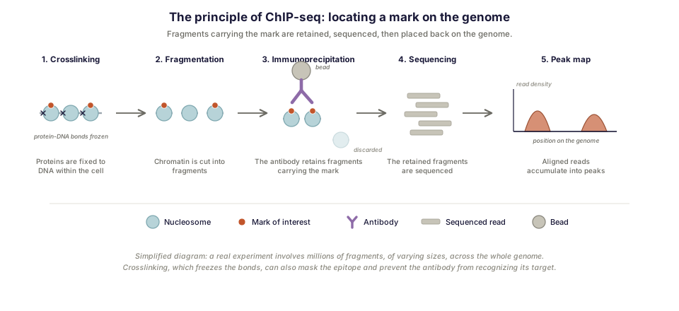
Chromatin immunoprecipitation followed by sequencing rests on a simple sequence of steps. Proteins are first fixed to DNA by chemical crosslinking, so as to freeze interactions as they existed in the cell. The chromatin is then fragmented, most often by sonication. Immunoprecipitation follows: an antibody directed against the mark or protein of interest captures the fragments carrying it, the others being removed by washing. The retained fragments are finally sequenced, and the resulting reads aligned to the reference genome.
The result is a map of read density along the genome. Where the mark was present, fragments accumulate and form a peak; elsewhere the signal stays at background level. This approach was introduced in 2007, applied from the outset both to mapping protein-DNA interactions in vivo [2] and to mapping histone methylations [1].
Narrow peaks, broad domains: two analytical regimes
One distinction conditions all interpretation, and it is too often neglected when designing the experiment.
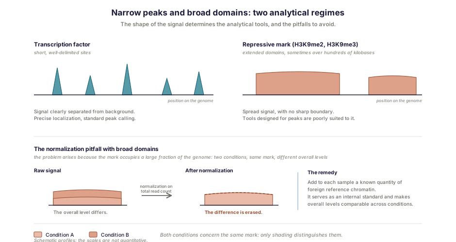
A transcription factor binds at short, well-delimited sites. Its signal therefore appears as a series of narrow peaks, clearly separated from background, easy to localize precisely.
Repressive histone marks behave quite differently. A mark such as H3K9me2 or H3K9me3 is not confined to a few sites: it covers broad domains, sometimes extending over hundreds of kilobases, and may concern a considerable fraction of the genome. The practical consequences are important. Peak-calling algorithms designed for narrow signals are poorly suited to these domains. And normalization becomes a problem in itself: when two conditions differ in the overall level of the mark, comparing signals normalized on total read count can erase precisely the difference one is trying to measure.
What makes or breaks an experiment
The ENCODE and modENCODE consortia, having found considerable differences in the way these experiments are conducted, scored and evaluated, established best-practice recommendations. These bear, in order, on antibody validation, experimental replication, sequencing depth, data and metadata reporting, and quality assessment [3]. That order is no accident.
The antibody is indeed the main point of failure. A reagent that recognizes its target poorly, or recognizes something else in addition, produces a perfectly plausible map (peaks, domains, a credible distribution) that is nonetheless wrong. No downstream analysis makes up for that defect, and nothing in the data signals it. Hence the requirement for prior validation, by independent controls.
Then come the experimental controls: an input control, corresponding to fragmented but non-immunoprecipitated chromatin, which gives the expected background; and, when comparing conditions liable to differ in the overall level of the mark, normalization by adding a known quantity of foreign reference chromatin.
There remain constraints intrinsic to the method, which the designers of the following approaches catalogued themselves: ChIP-seq suffers from weak signals, high background, epitope masking due to crosslinking — the very step that freezes interactions can render the target inaccessible to the antibody — and its low yields demand large numbers of cells [5]. This last constraint is often the most limiting in practice: when the cells of interest are rare (sorted hematopoietic stem cells are counted in thousands, not millions), it becomes an obstacle. That is what motivated the development of the methods taken up next.
From ChIP-seq to CUT&Tag: the shift under way
Another strategy has established itself over the past decade, and it inverts the logic. Rather than extracting and then sorting the fragments carrying the mark, the cutting enzyme is brought directly into contact with the target, in intact cells or nuclei.
The principle first took the form of CUT&RUN, in which a protein A fused to a micrococcal nuclease is guided by the antibody to the mark, then activated to cut DNA in the immediate vicinity [4]. CUT&Tag extends the idea by replacing the nuclease with a Tn5 transposase, which cuts and simultaneously inserts the sequencing adapters, doing away at a stroke with sonication, immunoprecipitation and most of library preparation. All the steps, from living cells to sequencing-ready libraries, fit in a single tube and are carried out in a day [5].
The gains are substantial and threefold. The signal-to-noise ratio is markedly better, since only what lies near the antibody is cut: the authors describe high-resolution libraries with exceptionally low background. That property has a direct economic consequence: in situ mapping requires only about one tenth of the sequencing depth demanded by ChIP [4]. And above all, the quantity of material needed falls, which opens access to rare samples, clinical specimens and sorted populations.
This last property has made practicable what ChIP-seq allowed only at prohibitive inefficiency: single-cell profiling. A recent review sums up the shift bluntly — conventional bulk approaches such as ChIP-seq require large cell populations, which limits their applicability to heterogeneous samples and tissues, whereas single-cell CUT&Tag and its variants allow high-resolution profiling of histone modifications and transcription factors in individual cells, extending single-cell epigenomics beyond chromatin accessibility alone [6]. Some of these variants use single-domain antibodies fused to the transposase, which makes it possible to profile several marks simultaneously within one cell — one more application of these single-domain antibodies, which we treat elsewhere.
Two qualifications are nevertheless in order. Using the Tn5 transposase brings CUT&Tag close to ATAC-seq, a technique that exploits the same enzyme’s preference for nucleosome-free regions in order to map chromatin accessibility — a different question from that of a specific mark. That kinship creates a risk of unguided adapter integration, independent of the antibody — a risk the method’s designers control explicitly by running the experiment with a non-specific antibody, which must then produce a landscape very poor in signal [5]. And ChIP-seq retains an advantage that is not slight: nearly twenty years of public data, reference datasets and tested recommendations, against which any new result can be compared.
The case: a tightened repression, and how to lift it
Work Inovarion contributed to shows what mapping a repressive mark makes it possible to establish, and the result runs against intuition.
Chronic myelomonocytic leukemia is a severe myeloid malignancy of the elderly, with a median age at diagnosis of 72, arising from a hematopoietic stem cell and classed among myelodysplastic/myeloproliferative neoplasms. The prognosis is poor — median survival is under three years in most series — the only curative option remains allogeneic transplant, difficult to carry out given age and comorbidities, and DNA hypomethylating agents provide only transient responses without eradicating the malignant clone [8].
The study’s starting point is a known observation: aging of hematopoietic stem cells is accompanied by a reorganization of heterochromatin, attested by alterations of the H3K9me2 and H3K9me3 marks — earlier work having shown that the NF-κB pathway controls H3K9me3 levels at intronic LINE-1 elements and hematopoietic stem cell genes [7]. The team therefore mapped these marks in patients’ stem cells and progenitors.
The result is unexpected. One might have expected a malignant clone to loosen repressive control of its genome. The opposite is observed: heterochromatin is indeed disorganized, but with an increase in the H3K9me2 mark, mainly at transposable elements, and a repression of immune and age-associated transcripts. The disease does not loosen its grip, it tightens it, and that tightening switches off immune pathways.
The therapeutic reasoning then follows a logic opposite to the usual one. If the problem is an excess of repression, what is needed is not to inhibit a function but to lift a silence. By combining hypomethylating agents with inhibitors of the G9A and GLP methyltransferases responsible for the H3K9me2 mark, the team reactivates retroelements and immune pathways. And the result that gives the approach its credibility is its selectivity: mutated cells are eliminated, while non-mutated stem cells are preserved.
This disease has also been approached, in other work Inovarion contributed to, from the angle of the transcriptome — two layers of reading on one and the same pathology, chromatin saying what is kept silent, the transcriptome what is actually expressed.
Limits and points of vigilance
Three pitfalls recur, and knowing them conditions the value of a project.
Antibody specificity remains the first, whatever the method used: guided-nuclease approaches depend on an antibody just as much as ChIP-seq does. Changing technique does not dispense with validating the reagent.
Normalization of broad domains is the second, particularly when the conditions compared differ in the overall level of the mark — the exact case of the study discussed above.
The third is logical, and it holds for any mapping. Observing that a mark occupies a region does not establish that it exerts an effect there, and co-occurrence is not causation. Establishing a function requires perturbing (by genome editing or pharmacological inhibition, as in the case above) and then observing the consequence, including on the transcriptome. It is the articulation of these layers, not any one of them, that produces a solid conclusion.
How Inovarion can support you
Inovarion takes on epigenomics projects from design through to interpretation: choice of method according to the nature and quantity of material available, experimental implementation of ChIP-seq as well as guided-nuclease approaches, validation of antibodies and controls, then bioinformatic analysis — peak calling, handling of broad domains, normalization suited to differences in overall level, and integration with transcriptomic data. It is this dual command, experimental and analytical, that separates an interpretable map from a plausible profile.
Publications
Field references
- Barski A, Cuddapah S, Cui K, et al. High-resolution profiling of histone methylations in the human genome. Cell, 2007;129(4):823-837. DOI
- Johnson DS, Mortazavi A, Myers RM, Wold B. Genome-wide mapping of in vivo protein-DNA interactions. Science, 2007;316(5830):1497-1502.
- Landt SG, Marinov GK, Kundaje A, et al. ChIP-seq guidelines and practices of the ENCODE and modENCODE consortia. Genome Research, 2012;22(9):1813-1831. DOI
- Skene PJ, Henikoff S. An efficient targeted nuclease strategy for high-resolution mapping of DNA binding sites. eLife, 2017;6:e21856. DOI
- Kaya-Okur HS, Wu SJ, Codomo CA, et al. CUT&Tag for efficient epigenomic profiling of small samples and single cells. Nature Communications, 2019;10:1930. DOI
- Wu J, Wahiduzzaman M, Yin P, et al. Advances in scCUT&Tag and computational analysis for single-cell gene regulatory element mapping. Briefings in Bioinformatics, 2026;27(1):bbag015. DOI
- Pelinski Y, Hidaoui D, Stolz A, et al. NF-κB signaling controls H3K9me3 levels at intronic LINE-1 and hematopoietic stem cell genes in cis. Journal of Experimental Medicine, 2022;219(8):e20211356.
Inovarion contribution
- Hidaoui D, Porquet A, Chelbi R, et al. Targeting heterochromatin eliminates chronic myelomonocytic leukemia malignant stem cells through reactivation of retroelements and immune pathways. Communications Biology, 2024;7(1):1555. Record → · DOI
updated July 2026