Classical genetics usually proceeds one gene at a time: a gene suspected of playing a role is inactivated, and the resulting phenotype is observed. This approach has been productive, but it runs into a limit of scale — the human genome contains roughly twenty thousand genes, and interrogating them one by one becomes impossible once the question turns into “among all these genes, which ones matter for this phenotype?”. CRISPR-Cas9 removed that obstacle by making it possible to inactivate thousands of genes in parallel within a single experiment. This is the principle of the genome-scale CRISPR screen, one of the most powerful tools in contemporary functional genomics. This article offers an overview: principle, readout strategies, formats, toolbox, interpretation, strengths and pitfalls.
From single gene to genome-scale screen
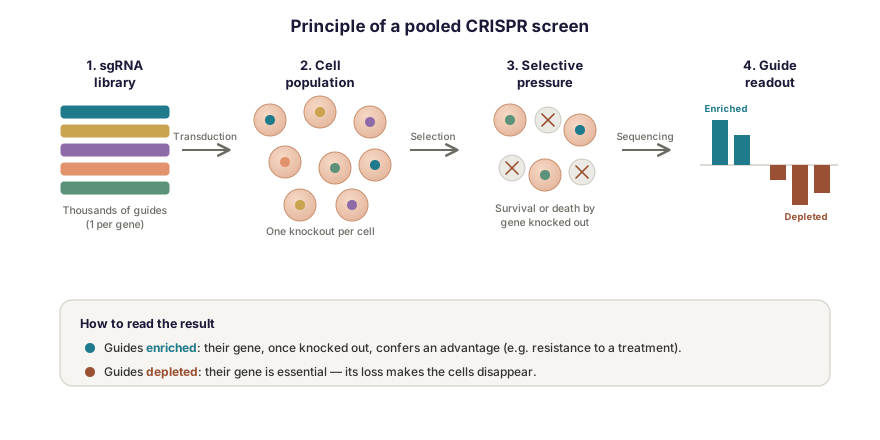
The CRISPR-Cas9 mechanism is now well established: a guide RNA (sgRNA) directs the Cas9 nuclease to a target sequence in the DNA, where it introduces a double-strand break. The cell’s repair of that break is often imperfect and generates small insertions or deletions that shift the reading frame and inactivate the gene — this is the knockout. Applied to a single gene, the mechanism allows its function to be studied. What is new about screening lies entirely in a change of scale: instead of a single sgRNA, a library of thousands of different sgRNAs is introduced into a cell population, so that each cell in principle receives one guide and carries the knockout of a different gene.
The result is a population in which, collectively, almost every gene is switched off — each in a subset of cells. All that remains is to apply a selective pressure and read out, by sequencing, which guides were favored or eliminated. The founding library of this approach, GeCKO, already targeted more than 18,000 human genes with close to 65,000 guides [1]; an analogous strategy was published simultaneously by another group [2].
Two readout strategies
What you are looking for determines how the screen is read. Two complementary logics coexist.
Dropout screens: finding essential genes
In a depletion screen, cells are simply allowed to proliferate over several generations. Cells that have lost a gene essential to their survival or division gradually disappear, so the corresponding guides become less and less abundant over time. Comparing sgRNA abundance at the start and end of the experiment identifies genes whose loss is deleterious — essential genes, or, in a tumor context, the vulnerabilities specific to a cancer type.
Positive selection screens: finding resistance genes
Conversely, a pressure can be applied that kills or blocks most cells: a drug, a pathogen, a stress. The few cells that survive are those whose knockout confers an advantage, such as resistance to treatment. The guides they carry then become strongly enriched. This type of screen is particularly useful for understanding resistance mechanisms, by identifying genes whose loss allows a cell to escape a treatment.
In both cases the information is carried by the relative abundance of sgRNAs, measured by high-throughput sequencing before and after selection: the screen turns a biological question into a comparison of counts.
Pooled or arrayed: two formats
How cells are physically organized defines two broad families of screens.
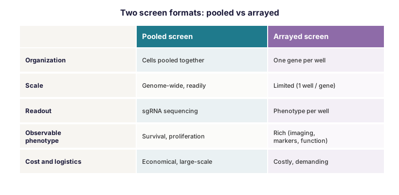
In the pooled format, all cells are mixed in a single vessel, each carrying its own knockout. The readout is exclusively by sgRNA sequencing: this is economical, massively parallel, and suited to genome-wide libraries. The trade-off is that the observable phenotype is limited to whatever affects cell abundance — survival, proliferation, the ability to pass a selection.
In the arrayed format, by contrast, each gene is targeted in a separate well of a plate. Every well can then undergo rich phenotyping: imaging, fluorescent markers, fine functional measurements. This format opens up questions inaccessible to the pooled approach, but it is markedly more expensive and more demanding to implement, which limits its scale.
Beyond knockout: the CRISPR toolbox
Knockout by double-strand break is only one of the available modalities. The same logic of guide-RNA-programmable targeting has been adapted into several variants, considerably widening the scope of screening.
Disabling the cutting activity of Cas9 — giving what is known as dCas9, for dead Cas9 — while preserving its targeting capacity allows modules to be attached that repress or activate the transcription of a gene without ever cutting the DNA: this is CRISPR interference (CRISPRi) and CRISPR activation (CRISPRa). These approaches make it possible to modulate expression finely rather than switch it off abruptly, and to interrogate non-coding regulatory regions as well. Other techniques still, such as base editing and prime editing, modify the sequence without inducing a double-strand break, with greater precision. The value of these variants is not merely technical: by avoiding DNA breaks they circumvent part of the toxicity associated with knockout screens, and make it possible to study essential genes that a complete knockout would simply render lethal.
What CRISPR screens are used for: main applications
The versatility of the approach explains the diversity of its uses. Several broad families of application have become established.
The first is the identification of therapeutic targets. By pinpointing genes whose loss is fatal to cancer cells but not to healthy ones, depletion screens reveal vulnerabilities specific to a tumor, each a potential target for new treatments. Mapping essential genes across cell lines is today an entire branch of oncology research.
The second, very active, is understanding resistance to treatment. By applying a drug as selective pressure, a screen identifies genes whose loss makes cells resistant or, conversely, sensitive — shedding light on escape mechanisms from chemotherapy, targeted therapy and immunotherapy, and suggesting combination strategies to circumvent them.
Screens also serve to map biological pathways and genetic interactions: by crossing the effects of many knockouts, functional networks are reconstructed and a gene of unknown function can be placed within an already characterized pathway. In infectious disease, they make it possible to identify the host factors a virus or bacterium requires to infect the cell — antiviral targets that are all the more attractive because the pathogen can hardly circumvent them. Finally, applied to immune cells, notably by CRISPRa activation in T lymphocytes, screens contribute to improving cell therapies by identifying the modifications that strengthen antitumor efficacy.
Reading and interpreting a screen
The raw result of a pooled screen is not a list of genes but a table of counts: how many times each sgRNA was read, before and after selection. Turning those counts into biological conclusions requires dedicated analysis. The reference tool in this area, MAGeCK, was designed precisely to rank guides, genes and pathways from such data: it normalizes the counts, compares conditions, aggregates the information from the different guides targeting a given gene, and controls the false discovery rate [3].
One point conditions the robustness of any conclusion: a screen “hit” is never proof, only a hypothesis. A gene that emerges as enriched or depleted must be validated individually — by repeating the experiment with individual guides, by checking that reintroducing the gene restores the phenotype, or by cross-checking against other approaches. A well-designed screen produces a ranked list of candidates; it is the validation work that turns it into a discovery.
Strengths, limits and pitfalls
The strength of the CRISPR screen is that it is systematic and unbiased: it requires no prior hypothesis about the genes involved, interrogates the genome as a whole, and establishes causality through loss of function. But that power comes with pitfalls that must be handled.
Off-target effects, cuts at unintended sites in the genome, can generate false signals, even though better guide design has reduced them. The knockout may also be incomplete or mosaic: some insertions or deletions preserve the reading frame and leave a partially functional gene. A subtler difficulty lies in the toxicity of double-strand breaks themselves: in primary or non-transformed cells, these breaks trigger a p53 response that can distort the screen by disadvantaging cells simply because they have sustained many cuts, regardless of the gene targeted. Finally, the reliability of a screen rests on library coverage: enough cells carrying each guide are needed for variations in abundance to be statistically interpretable.
Design and analysis make the result
A successful screen is decided as much upstream, at the design stage, as downstream, at analysis. The choice of library, the coverage aimed for, the number of replicates, positive and negative controls, the duration of selection: all are decisions that determine the quality of the result before the first sequencing run. Downstream, the bioinformatic handling of counts and rigorous control of false positives matter just as much. It is this dual requirement, experimental and analytical, that separates a usable screen from an accumulation of uninterpretable data.
For the technical workflow of CRISPR-Cas9 functional genomics (editing, RNA interference, loss-of-function validation) see our dedicated page CRISPR & functional genomics.
How Inovarion can support you
Inovarion supports functional genomics projects using CRISPR-Cas9 screens, from the choice of strategy through to the interpretation of results: selecting the format (pooled or arrayed) and the readout logic (depletion or enrichment), choosing between knockout, CRISPRi/CRISPRa or break-free editing, designing the library and its controls, then bioinformatic analysis and candidate validation. The aim is always the same: to match the screening strategy to the biological question, and to ensure the rigour that turns a list of hits into solid knowledge.
Further reading
- Shalem O, Sanjana NE, et al. Genome-Scale CRISPR-Cas9 Knockout Screening in Human Cells. Science, 2014;343(6166):84-87. DOI
- Wang T, Wei JJ, Sabatini DM, Lander ES. Genetic screens in human cells using the CRISPR-Cas9 system. Science, 2014;343(6166):80-84. PubMed
- Li W, Xu H, Xiao T, et al. MAGeCK enables robust identification of essential genes from genome-scale CRISPR/Cas9 knockout screens. Genome Biology, 2014;15(12):554. PubMed
updated July 2026