Between biochemistry on purified protein and animal experimentation, disease modeling long lacked a tier: the patient’s own human cell, carrying their mutation, and able to become precisely the cell type affected. Immortalized lines offer convenience but drift away from the tissue of origin; primary cells are faithful but rare and poorly proliferative; animal models integrate the whole organism at the cost of differences between species.
Induced pluripotent stem cells, iPSCs, occupy that missing tier. Reprogrammed from an adult cell of the patient, they multiply indefinitely and differentiate toward the cell type of interest, keeping the genome and therefore the mutation of that person. This article sets out the principle of these models, what they make it possible to discover — through a case where the answer was inaccessible otherwise — and the limits worth knowing before committing a project to them.
What iPSCs bring, and at which tier
Three properties combine, and it is their combination that makes the model valuable.
The material is human, which removes the divergence between species — the obstacle we describe in relation to preclinical models, where a candidate designed against a human protein may fail to recognize its murine homolog. It then carries the patient’s genome, and so their mutation in its own genetic context, where an overexpression system places the mutant protein in an artificial environment. It is finally differentiable toward the cell type actually diseased: megakaryocyte, cardiomyocyte, neuron, macrophage, endothelial cell. Studying a platelet disorder in megakaryocytes rather than in a line of convenience is not a refinement; it is often the condition for the phenotype to appear at all.
What these models do not replace must be said as well. A cell in culture, however well differentiated, has neither circulation, nor a complete immune system, nor interactions between organs. Questions of pharmacokinetics, systemic toxicity or integrated phenotype remain the province of in vivo work. The logic is not substitution but complementarity — each tier answering the questions proper to it.
The principle: reprogram, then differentiate
The founding demonstration dates from 2006. By introducing four transcription factors (Oct3/4, Sox2, c-Myc and Klf4) into mouse fibroblasts, embryonic or adult, a Japanese team obtained cells showing the morphology and growth properties of embryonic stem cells, expressing their marker genes, and forming, after grafting, tumors containing tissues derived from all three germ layers. One detail catches the eye: the factor Nanog, expected to be indispensable, proved dispensable [1].
The following year the same approach was transposed to humans, from adult dermal fibroblasts [2]. The motivation was explicit and it illuminates the whole point of the technique: human embryonic stem cells raised ethical questions, and above all it was difficult to obtain any that were patient- or disease-specific. Direct reprogramming lifts that obstacle — which is what grounds the use of iPSCs as disease models.
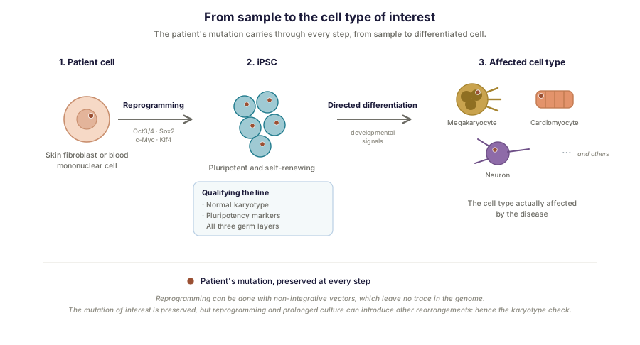
The route therefore has two stages. A somatic cell from the patient — skin fibroblast, blood mononuclear cell — is reprogrammed into a pluripotent cell. That cell is then directed, by differentiation protocols reproducing developmental signals, toward the cell type to be studied.
Generating and validating a line: what it involves
An iPSC line is not a reagent you order: it is a biological object that has to be qualified. Work Inovarion contributed to gives a concrete view of this, through the description of a line derived from a patient with a disease linked to the STING protein [6]. Mutation of the STING1 gene leads to constitutive activation of the protein, and thereby to a severe vasculopathy, sometimes accompanied by a lupus-like picture. The line was established from a patient carrying a rare heterozygous variant, c.463G>A leading to the p.V155M substitution.
Two points of that description matter. Reprogramming was carried out by non-integrative viral transduction: the reprogramming factors are delivered without inserting into the genome, which avoids introducing genomic modifications liable to confound later interpretation. And validation rests on three complementary checks: a normal karyotype, expression of pluripotency markers, and effective capacity for differentiation toward the three germ layers. This triad forms the minimum basis for qualifying a line; omitting it amounts to working on material you do not know to be what you think it is.
The decisive case: when the model reveals what assay cannot see
Work Inovarion contributed to illustrates strikingly what these models make it possible to establish, and why no other approach got there.
Bernard-Soulier syndrome is a severe bleeding disorder combining moderate to severe thrombocytopenia, abnormally large platelets and platelet dysfunction. It results from biallelic mutations of the GP1BA, GP1BB or GP9 genes, which encode the subunits of the GPIb-IX-V complex, the platelet receptor for von Willebrand factor. The case reported here fell outside that frame: a patient carrying a previously undescribed heterozygous mutation, GP1BA p.N103D, presenting with moderate macrothrombocytopenia [7]. How could a single mutated copy produce a phenotype?
iPSCs were derived from this patient, then differentiated into megakaryocytes. And this is where the result becomes instructive, because it is negative where one expected it to be positive. The mutation affects neither megakaryocytic differentiation nor expression of the GPIb-GPIX complex at the cell surface. Only affinity for von Willebrand factor is reduced. In other words, a protein assay or surface cytometry would have shown nothing abnormal in the production or the trafficking of the receptor. As for sequencing, it does identify the variant — that is how it was found — but says nothing of its functional consequence.
The defect lies elsewhere, and is of another nature: it is a gain of signaling. The mutation induces increased signaling, independent of von Willebrand factor, running through the so-called “outside-in” signaling of integrin αIIbβ3. Preactivation of that integrin is observed, together with increased stress fiber formation, linked to overactivation of the RhoA pathway, most likely consequent on increased phosphorylation of the kinase SRC at residue Y419, downstream of GPIbα. The functional consequence is a profound defect in proplatelet formation after adhesion on fibrinogen — that is, a defect in platelet production itself. The mutation therefore affects two distinct aspects: the receptor’s affinity for its ligand, and thrombopoiesis itself.
This result was accessible only to this kind of model. Human megakaryocytes were needed, carrying the mutation in its genetic context, and placed in a functional situation of adhering and then extending proplatelets, for the phenotype to manifest and its mechanism to be dissected. A purified protein would have said nothing about intracellular signaling; an immortalized line, which reproduces proplatelet formation poorly, would not have restored this phenotype.
It is worth noting in passing that GPIbα and von Willebrand factor are precisely the two proteins at the center of the humanized mouse model of von Willebrand disease and of the single-domain antibodies directed against that factor, which we treat elsewhere. One and the same molecular pair, approached through three distinct technologies — humanized animal model, VHH reagent, iPSC cell model — each answering a question the others could not settle.
The isogenic control: the major methodological asset
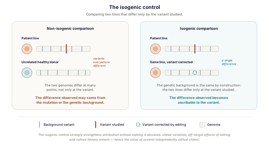
One objection shadows any study comparing a patient’s line with that of a healthy control: the two individuals differ not only by the mutation but by their entire genome. Any difference observed is therefore potentially confounded by genetic background, and attribution of the phenotype to the variant remains fragile.
The answer is the isogenic control. By genome editing, the mutation is corrected in the patient’s line, or introduced into a healthy line. You then have two lines that differ only by the variant studied — identical by construction, if not in strict terms, for the reasons given below. This design considerably strengthens attribution: what distinguishes the two lines becomes very plausibly ascribable to the mutation. It does not establish it absolutely, however, since clonal variation, off-target effects of editing and each line’s own culture history remain, which is why it is worth working on several independently edited clones.
This is where genome editing tools, whose principles we describe in relation to CRISPR-Cas9 screens, become indispensable not as an object of study but as an instrument of experimental rigor.
Limits and points of vigilance
These models have documented weaknesses, and knowing them conditions the quality of a project.
The first is variability between lines. Two lines from two donors, or even two clones from the same donor, can behave differently, owing to genetic background, the clone selected, or culture history. The remedy lies in two principles: working on several independent clones, and using isogenic controls as far as possible.
This limit is nevertheless subject to an instructive reversal. iPSCs have traditionally served to model monogenic diseases, where a highly penetrant variant produces a robust phenotype detectable across a small number of lines — the two cases described above are of that kind. Recent advances in scaling and standardization now allow systematic comparisons across many donors, which opens the field of complex diseases, driven by multiple variants of small individual effect and therefore requiring population-scale designs to resolve genotype-phenotype relationships [3]. In such designs, variability between lines ceases to be noise to be controlled and becomes the very signal under study. The two strategies are therefore not opposed: the isogenic control isolates the effect of a single variant, while donor panels map the joint effect of many variants. The choice depends, once again, on the question asked.
The second is the maturity of the cells obtained. In vitro differentiation protocols frequently produce cells whose profile remains immature, closer to the fetal stage than to adult tissue. The limit is particularly discussed for cardiomyocytes and neurons, and it calls for caution in interpreting diseases with a late-onset component.
Then come the reproducibility of protocols, sensitive to reagent lots and to details of handling, and the absence of tissue context: a culture of isolated cells reproduces neither the architecture of a tissue nor the dialogue between cell types. It is this last gap that organoids, cocultures and organ-on-chip systems seek to fill — and it is also what justifies retaining in vivo experimentation for the questions that require it.
A place strengthening within the regulatory landscape
This growing relevance meets a regulatory trajectory. The Food and Drug Administration announced in April 2025 its intention to phase down the requirement for animal testing in safety assessment, drawing notably on organoids and human cell systems [4]. The European Commission adopted, on 1 June 2026, a roadmap toward phasing out animal testing for chemical safety assessments, together with an investment effort in non-animal approaches, including in vitro technologies [5].
The nuance we stress in relation to preclinical models holds here too, and it would be imprudent to forget it: these texts bear first on the regulatory assessment of safety, not on fundamental research or disease modeling. But the investment and qualification effort they set in motion benefits human cell models directly, of which iPSCs are one of the pillars.
How Inovarion can support you
Inovarion takes on the full chain of iPSC-derived models: deriving lines from patient cells and qualifying them (karyotype, pluripotency markers, differentiation capacity), directed differentiation toward the relevant cell type, building isogenic controls by genome editing, then functional phenotyping and analysis. This expertise dovetails with our work on in vivo preclinical models and on high-throughput approaches, so that the choice of modeling tier can follow the question asked rather than the tool available.
Publications
Field references
- Takahashi K, Yamanaka S. Induction of pluripotent stem cells from mouse embryonic and adult fibroblast cultures by defined factors. Cell, 2006;126(4):663-676. PubMed
- Takahashi K, Tanabe K, Ohnuki M, et al. Induction of pluripotent stem cells from adult human fibroblasts by defined factors. Cell, 2007;131(5):861-872. DOI
- Adams HHH, et al. Bridging population and cell: modelling complex diseases with human induced pluripotent stem cells. European Journal of Human Genetics, 2026;34:741-748. DOI
- U.S. Food and Drug Administration. Roadmap to Reducing Animal Testing in Preclinical Safety Studies, April 2025.
- European Commission. Roadmap towards phasing out animal testing for chemical safety assessments, communication C(2026)3497, adopted 1 June 2026.
Inovarion contributions
- Barnabei L, Castela M, Banal C, Lefort N, Rieux-Laucat F. Generation of an iPSC line (IMAGINi011-A) from a patient carrying a STING mutation. Stem Cell Research, 2021;50:102107. Record → · DOI
- Lordier L, Di Buduo CA, Kauskot A, et al. Increased RhoA pathway activation downstream of αIIbβ3/SRC contributes to heterozygous Bernard Soulier syndrome. Haematologica, 2025;110(7):1596-1609. Record → · PubMed
updated July 2026