Single-cell sequencing presents itself as an analytical technique. It is first of all a technique of preparation. Between sampling and the first readout, cells have to be separated from one another, kept alive, and delivered intact to capture — and each of these steps decides what the experiment will be able to show.
This note sets out what dissociation really does to a tissue, why some populations disappear without leaving a trace, the decisions that commit everything else, and what analysis does not recover. Two pieces of work Inovarion contributed to serve as illustrations, one on blood, the other on solid tissue.
What dissociation does to a tissue
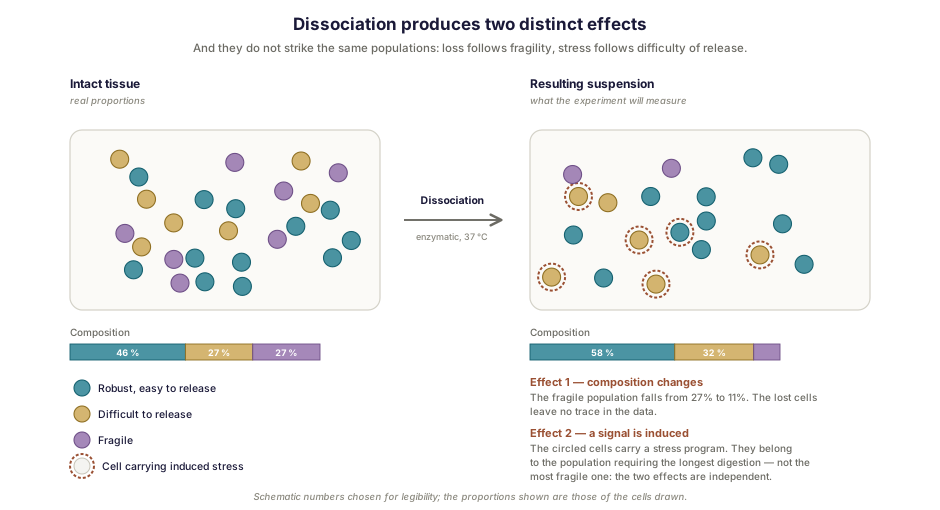
Two effects are superimposed, and it matters not to confuse them, since they call for different remedies.
It alters composition
Not all cells are released with the same ease. Depending on the enzyme, the duration, the temperature and the agitation, some populations are over-represented, others depleted, and the most fragile simply destroyed. The proportions observed in the data are therefore not those of the tissue. A systematic assessment of these biases, covering both dissociation and storage conditions, compared whole-cell and isolated-nucleus workflows [2].
And it manufactures a signal
The second effect is more troubling, because it translates not into a loss but into a production: dissociation induces in a proportion of the cells a stress response program [1]. The artifact is therefore not measurement noise but a real transcriptional response, triggered by the treatment itself, which makes it interpretable, and thereby misleading.
The mechanism is well established. Classical enzymatic dissociation imposes incubation at 37 °C, a temperature at which the cell transcribes actively and therefore reacts to what is being done to it. One team exploited that property in reverse, using a cold-active protease purified from a psychrophilic microorganism: the entire procedure takes place at 6 °C or below, a temperature at which the mammalian transcriptional machinery is largely inactive, which amounts to “freezing” expression profiles as they were in vivo. Compared with 37 °C protocols on more than twenty thousand cells, this approach strongly reduces expression artifacts [3]. The principle was subsequently transposed to solid tumors, where it limits the collagenase-associated stress responses [4].
One detail completes the artifact’s capacity to mislead. It does not affect cells at random: the authors find that a subpopulation of osteoblasts, cells difficult to release from the tissue, carries the same stress signature as the affected stem cells [1]. When the artifact is thus correlated with cell type, it can be read as a property of the population rather than as a trace of the protocol.
Beyond the choice of protease, the surest remedy remains to plan from the design stage the means of distinguishing artifact from biology: compared conditions, controls treated differently, or verification on a modality that does not dissociate.
The difficult case: granulocytes
The clearest illustration of these difficulties comes from a population that most datasets simply ignore.
In studies conducted on droplet platforms, neutrophils are completely absent or very rarely represented. Yet cytometry establishes that they make up 10 to 20% of the leukocytes of lung tumor tissue and adjacent tissue, across a series of 63 samples. This under-representation is therefore technical in origin, and its causes compound: these are fragile cells, with a circulating half-life of seven to ten hours in humans, particularly sensitive to handling, and they express an exceptionally low quantity of messenger RNA molecules [5]. To these properties are added a high content of RNases — low messenger RNA content and abundance of nucleases being precisely what makes their profiling difficult [6] — as well as loss on cryopreservation.
What this study did with that difficulty is the most convincing argument in favor of careful preparation. The authors integrated close to 1.3 million cells from 556 samples and 318 patients, adding their own dataset designed to capture cells with low RNA content. That analysis revealed subpopulations of resident neutrophils acquiring new functional properties in the microenvironment, and the gene signature derived from them turned out to be associated with failure of anti-PD-L1 treatment [5].
In other words, the population that sample preparation was losing was the one carrying the clinical signal.
Two cases, two matrices
Work Inovarion contributed to tackled this same population in a hematological context. The immature granulocytes of patients with chronic myelomonocytic leukemia were detected and quantified by flow cytometry, then subjected to bulk and single-cell sequencing, which revealed their pro-inflammatory profile — CXCL8 being the most abundantly secreted cytokine. Accumulation of these cells proved a powerful and independent adverse prognostic factor [8]. Single-cell sequencing of granulocytes is therefore not out of reach; it presupposes treating fragility as a design constraint, not as a hazard. We return to the sorting of this population in our note on cytometry panels, and to the choice between bulk and single-cell sequencing.
Blood, however, dispenses with dissociation. A second piece of work Inovarion contributed to illustrates the case of solid tissue: memory B lymphocytes specific for a viral antigen were isolated from the spleens of organ donors, in people vaccinated more than forty years earlier, then characterized by single-cell sequencing [9]. Two difficulties proper to tissue are then added: dissociation itself, and the sorting of a population whose frequency is minute — the second making sense only if the first has not destroyed what is being sought.
The decisions that commit everything else
Whole cells or isolated nuclei
When the cell does not survive dissociation, the nucleus may withstand it. Nucleus isolation thus makes it possible to approach frozen tissues or cell types that are too fragile. The price is known: the cytoplasmic transcriptome is lost, and the profiles obtained are not directly comparable to those of a whole cell — which is why assessments systematically compare the two workflows [2].
Fresh, cryopreserved or fixed
Fresh processing remains the reference where possible, but it imposes a heavy logistical constraint. Cryopreservation permits storage, at the cost of selective loss — critical for granulocytes, which do not withstand it.
This is where the field has shifted recently. Methods based on fixation aim to preserve RNA integrity after sampling, and a multisite assessment showed that they retain fragile granulocyte populations better than fresh processing [7]. That same study offers an illustration of the principle set out above: in order to have an independent reference, the authors characterized their cells with a twenty-one-color cytometry panel — a modality that does not share the steps they were seeking to evaluate. And the consequence goes beyond the bench: since a fixed sample can be stored and transported, distributed collection and centralized processing become possible. A population that survives neither transport nor freezing made a multicenter study impracticable; fixation lifts that obstacle. The choice of preservation mode is therefore a project design decision as much as a technical one.
Viability, doublets, ambient RNA
Three indicators are monitored routinely, and each betrays a defect of preparation. Degraded viability signals overly aggressive dissociation and releases RNA into the medium. This ambient RNA is then captured indiscriminately and attributed to cells that were not expressing it. Doublets, finally, result from a suspension that is too concentrated or poorly dissociated; a comparison of methods reports up to about 20% of them on one of the chemistries evaluated [6].
What analysis does not recover
Analysis has real tools at its disposal. Ambient RNA is estimated and subtracted, doublets are detected and removed, low-complexity cells are filtered out. These corrections are useful and it would be absurd to forgo them.
They nevertheless have two limits that must be known before sampling.
The first is that an absent population cannot be reconstituted. If the granulocytes did not survive, no statistical treatment will make them appear, and nothing in the sequencing data alone will signal their absence: absence does not declare itself. It is detected, on the other hand, by external comparison — it was precisely by setting their datasets against cytometry, which gave 10 to 20% neutrophils, that the authors of the lung atlas established the under-representation. Measuring composition by a second modality is therefore the simplest remedy.
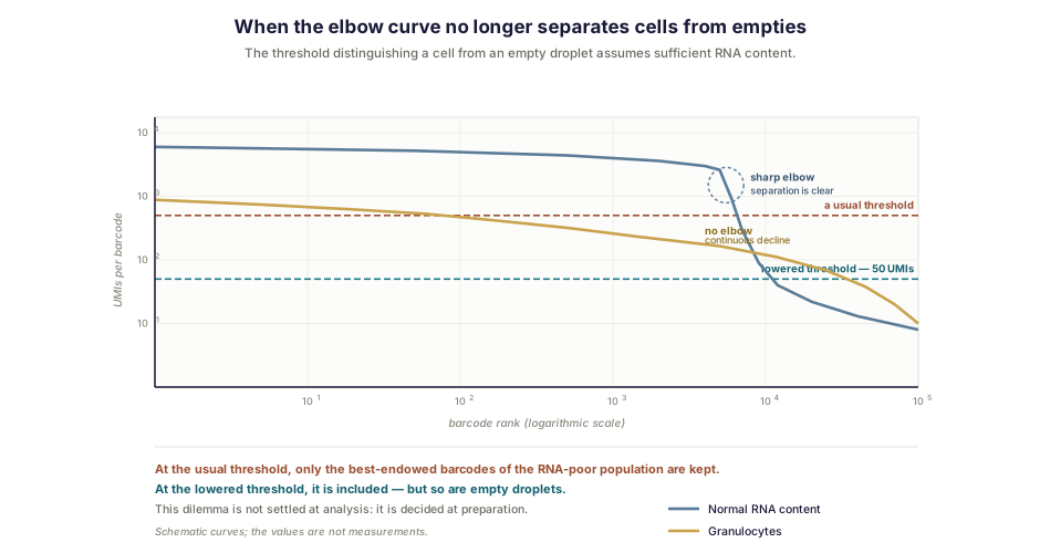
The second is subtler. Filtering thresholds are calibrated on cells with normal RNA content. On a sample rich in granulocytes, the elbow curve, which usually separates real cells from empty droplets without ambiguity, ceases to show a sharp separation, precisely because these cells express little. That comparison of methods had to lower the threshold to fifty genes and fifty unique molecular identifiers so as not to exclude them [6], which amounts to admitting noise in order not to lose the signal. No setting removes this dilemma: it is settled upstream, by the mode of preparation.
There is finally a response that consists not of dissociating better but of not dissolving the tissue at all. Spatial transcriptomics, which we treat elsewhere, measures expression on the section itself and therefore escapes the dissociation artifact by construction, at the cost of a resolution that, depending on the technique used, ranges from several cells per position to the micrometer scale.
Before sampling
Six questions, in this order, avoid most disappointments.
Which populations do you want to see, and which one carries the hypothesis? Are those populations reputed to be fragile or RNA-poor? Does logistics permit immediate processing, or must the sample be preserved, and if so, does the target population withstand freezing? Is the dissociation protocol suited to the tissue, and its duration reduced to what is necessary? Does the design include the means of distinguishing a preparation artifact from a biological signal? And have the analysis thresholds been set in knowledge of the populations expected, rather than by default?
How Inovarion can support you
Inovarion takes on the complete chain upstream of sequencing: designing the preparation protocol according to the populations targeted and the project’s logistical constraints, tissue dissociation, cell sorting, viability and quality controls, then bioinformatic analysis — doublet detection, ambient RNA correction, threshold calibration according to the populations expected. Our teams have carried out this work on populations reputed to be difficult, from the immature granulocyte to the extremely rare memory B lymphocyte, which presupposes treating preparation as part of the experiment and not as its preliminary formality.
Publications
Field references
- van den Brink SC, Sage F, Vértesy Á, et al. Single-cell sequencing reveals dissociation-induced gene expression in tissue subpopulations. Nature Methods, 2017;14(10):935-936. PubMed
- Denisenko E, et al. Systematic assessment of tissue dissociation and storage biases in single-cell and single-nucleus RNA-seq workflows. Genome Biology, 2020;21(1):130. DOI
- Adam M, Potter AS, Potter SS. Psychrophilic proteases dramatically reduce single-cell RNA-seq artifacts: a molecular atlas of kidney development. Development, 2017;144(19):3625-3632. PubMed
- O’Flanagan CH, Campbell KR, Zhang AW, et al. Dissociation of solid tumor tissues with cold active protease for single-cell RNA-seq minimizes conserved collagenase-associated stress responses. Genome Biology, 2019;20(1):210. DOI
- Salcher S, Sturm G, Horvath L, et al. High-resolution single-cell atlas reveals diversity and plasticity of tissue-resident neutrophils in non-small cell lung cancer. Cancer Cell, 2022;40(12):1503-1520.e8. PubMed
- Hatje K, Schneider K, Danilin S, et al. Comparison of single-cell RNA-seq methods to enable transcriptome profiling of neutrophils in clinical samples. Cell Reports Methods, 2025;5(9):101173. PubMed
- Kolling FW IV, Podnar JW, Wilkins O, et al. Multisite assessment of methods for cell preservation upstream of single-cell RNA sequencing. Journal of Biomolecular Techniques, 2026;37(2):9-27. DOI
Inovarion contributions
- Deschamps P, Wacheux M, Gosseye A, et al. CXCL8 secreted by immature granulocytes inhibits WT hematopoiesis in chronic myelomonocytic leukemia. The Journal of Clinical Investigation, 2024;134(22):e180738. Record → · DOI
- Chappert P, Huetz F, Espinasse MA, et al. Human anti-smallpox long-lived memory B cells are defined by dynamic interactions in the splenic niche and long-lasting germinal center imprinting. Immunity, 2022;55(10):1872-1890.e9. Record → · PubMed
updated July 2026