For a long time cancer research concentrated on the tumor cell itself: its mutations, its proliferation, its capacity to invade tissues. But a tumor is not a homogeneous mass of cancer cells. It is a living, complex tissue, in which malignant cells coexist with blood vessels, a supporting stroma and, above all, a large number of immune cells. This ensemble, the tumor immune microenvironment, is today recognized as a major determinant of how the disease evolves and of the response to treatment.
This shift of perspective has very concrete consequences. It is now known that the nature of a patient’s immune reaction against their own tumor can predict the risk of recurrence with an accuracy that rivals, and even exceeds, that of classical anatomical criteria. This article explains what the tumor immune microenvironment is, why it has become central in oncology, and by which experimental methods it is characterized.
What is the tumor immune microenvironment?
The tumor immune microenvironment designates all the immune cells present within the tumor and at its periphery, together with the tissue context in which they operate. It includes different populations of T lymphocytes — notably CD3+ T cells (a marker of all T lymphocytes) and cytotoxic CD8+ T cells, capable of killing tumor cells directly — but also B lymphocytes, macrophages, dendritic cells and NK cells, not forgetting the stroma and the vascular network that structure this milieu.
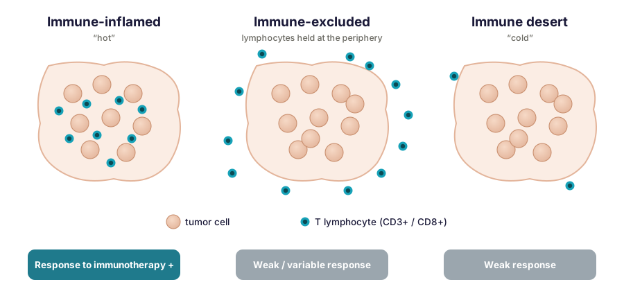
The composition of this infiltrate varies enormously from one tumor to another, and that is what makes it valuable clinical information. Two profiles are classically contrasted. So-called “hot” tumors are heavily infiltrated by active T lymphocytes: the immune system recognizes the tumor and attempts to control it. “Cold” tumors, by contrast, are little or not infiltrated; they largely escape immune surveillance. Between these two extremes lies a whole continuum.
This distinction is not merely a convenience of language: it covers profound differences of biological behavior. A hot tumor is generally associated with a better prognosis and responds better to immunotherapies, in particular to immune checkpoint inhibitors (such as anti-PD-1/PD-L1), which release the brakes on the lymphocyte response. A cold tumor often resists these treatments. Understanding why a tumor is hot or cold — and, in time, knowing how to “warm up” cold tumors — is one of the great objectives of contemporary immuno-oncology.
By taking in the spatial distribution of lymphocytes, and no longer their number alone, three profiles can be distinguished more finely along this continuum. The immune-inflamed phenotype (“hot”) shows abundant infiltration of CD8+ T lymphocytes in contact with tumor cells. The immune-excluded phenotype is characterized by lymphocytes present in numbers but held in the peripheral stroma, unable to penetrate the heart of the tumor. The immune-desert phenotype is defined by a scarcity of lymphocytes both in the tumor and in the surrounding stroma. This spatial reading has direct clinical bearing: inflamed tumors generally respond better to checkpoint blockade, whereas excluded and desert phenotypes are often little or not sensitive to it. Above all it underlines that the decisive information is not only “how many lymphocytes?”, but “where are they?”.
Immune contexture and its prognostic value
For decades, estimating the prognosis of a cancer rested on the TNM classification, which describes the anatomical extent of the disease: tumor size (T), nodal involvement (N), presence of metastases (M). These criteria remain fundamental, but they ignore an essential dimension: the host’s immune reaction.
The idea that this reaction carries prognostic value was established by a founding paper published in Science in 2006, which showed that in colorectal cancer the density and location of infiltrating T lymphocytes predicted clinical outcome, and proved a better predictor of survival than classical histopathological criteria [1]. The field was subsequently formalized under the term “immune contexture”: the type, density, location and functional organization of immune cells within the tumor [2].
It is on this foundation that the Immunoscore® was developed, a standardized test owned by INSERM and licensed to Veracyte, which quantifies by digital pathology the densities of CD3+ and CD8+ T lymphocytes in the tumor core and at its invasive margin. Inovarion has contributed to international validation studies of this type of biomarker in colon cancer, including a multicenter study of the score’s predictive value at stage III, for survival as well as for response to chemotherapy [5]. The international consensus validation study, coordinated by the Society for Immunotherapy of Cancer and published in The Lancet in 2018, covered tissues from 3,539 patients with stage I-III colon cancer, of whom 2,681 were retained in the analyses after quality control. It demonstrated high inter-site reproducibility and strong prognostic value: patients with a high Immunoscore showed a markedly lower five-year risk of recurrence than those with a low Immunoscore, and this association remained independent of age, sex, T stage, N stage and microsatellite instability [3].
In other words, measuring the immune reaction brings prognostic information that does not reduce to that of the anatomical stage. It opens the way to a classification of tumors that finally incorporates the immune dimension.
How an immune score is actually built
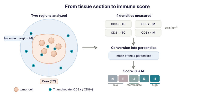
Behind this principle lies a precise and reproducible methodology. The score rests on two markers, CD3+ and CD8+, quantified in two distinct regions: the tumor core and the invasive margin, that is, the zone of about one millimeter centered on the invasion front, where the tumor meets healthy tissue. This gives four density measurements (two markers × two regions), expressed in cells per square millimeter.
These four densities are then converted into percentiles relative to a reference population, and averaged. In published implementations, the result is translated into a score running from I0 to I4 according to the number of measurements exceeding a high threshold, then grouped into three categories: low, intermediate or high. This conversion into percentiles is what allows samples to be compared with one another in a standardized way, and it is the robustness of that procedure that makes high reproducibility possible from one center to another — an indispensable condition for clinical use.
Beyond prognosis: guiding treatment
The value of characterizing the immune microenvironment goes beyond estimating risk. The degree and nature of immune infiltration directly influence the response to treatment. The level of lymphocyte infiltration is thus one of the major determinants of response to immunotherapies: tumors rich in CD8+ T lymphocytes are more likely to respond to checkpoint inhibitors. Immune characterization therefore becomes a decision-support tool, for identifying patients likely to benefit from one therapeutic strategy or another.
This translational reading, from bench to clinical decision, is at the heart of the approach: quantifying the immune environment finely means giving oneself the means to personalize care.
How the immune microenvironment is characterized
No single technique suffices to describe so rich a milieu. Several complementary approaches are combined, from whole tissue down to the single cell — a range Inovarion’s teams deploy daily.
Immunohistochemistry and digital pathology
The first step often consists of visualizing and counting immune cells directly on the tissue section. Immunohistochemistry (IHC) specifically labels the populations of interest — CD3, CD8 and many others. Digital pathology then takes over: by automated image analysis, it quantifies cell density in a standardized and reproducible way, where a pathologist’s visual appraisal remains subjective and difficult to harmonize across centers — a gap that a multi-institutional evaluation measured by comparing pathologists’ assessment with the Immunoscore [6]. This automated quantification is what allowed a biological observation to be turned into a reliable clinical test.
Quality control, guarantor of reliability. The value of an immune characterization is worth no more than the rigor of its execution. Several control points are decisive: validation and specificity of the antibodies used in IHC and multiplex; standardization of sample preparation (fixation, embedding, section thickness); definition of explicit and reproducible positivity thresholds; and monitoring of reproducibility between lots, between observers and between centers. It is this methodological framework, invisible in the final result but decisive, that separates a usable measurement from uninterpretable data, and on which Inovarion founds the reliability of its analyses.
Widening the panel of markers
CD3+ and CD8+ T lymphocytes are only the most studied part of a far wider ecosystem. To draw a complete portrait of the microenvironment, other markers are brought in, each revealing a particular functional population. The FoxP3 marker identifies regulatory T cells (Tregs), which exert an immunosuppressive effect and can restrain the antitumor response. The CD68 and CD163 markers distinguish tumor-associated macrophages, some subpopulations of which on the contrary favor tumor progression. The Ki67 marker reports on cell proliferation, while PD-1 and its ligand PD-L1 signal engagement of the immune checkpoints — information directly relevant to anticipating response to immunotherapies. Combining these markers, in particular by multiplex immunofluorescence, allows several populations to be analyzed on a single section and restores the real balance between pro- and antitumor forces.
Single-cell and spatial approaches
Counting cells does not always suffice: the aim is also to identify subpopulations finely and to understand their organization in space. Single-cell RNA sequencing reveals the diversity of cell states present in the tumor, well beyond what IHC distinguishes. Spatial transcriptomics adds the dimension of location, associating expression profile with position in the tissue. Spectral flow cytometry and mass cytometry, for their part, allow dozens of markers to be characterized simultaneously on each cell. Together, these methods draw a map of the immune infiltrate of a richness inaccessible to conventional techniques.
That richness has a downside: the analytical challenge. These methods impose technical choices that condition the quality of the results. Single-cell requires dissociating the tissue, which by construction destroys spatial organization; spatial approaches preserve it but often with lower resolution or molecular depth — a trade-off to be settled according to the biological question. Downstream, the volume of data is considerable and unusable without a rigorous bioinformatic pipeline: quality control, normalization, grouping of cells (clustering), cell type annotation, and, for spatial transcriptomics, deconvolution of signals when several cells mix within a single measurement spot. Characterizing the microenvironment is therefore as much bench work as computational analysis — a dual competence at the heart of what Inovarion does.
Studying the microenvironment at the scale of metastases
One and the same patient may carry several metastases whose immune microenvironments differ radically. Studying that heterogeneity, and its evolution over time under pressure from the immune system, sheds light on the mechanisms of tumor escape and progression.
Work Inovarion contributed to illustrates the power of this approach. A study published in Cancer Cell in 2018 quantified the immune populations of 603 metastases and primary tumors, on whole slides, in 222 patients with colorectal cancer. It revealed a deeply heterogeneous immune infiltrate from one lesion to another: small metastases frequently showed a low Immunoscore, while a high Immunoscore was associated with a smaller number of metastases [7]. The conclusion is strong: each metastasis behaves, immunologically, as a disease in its own right, and a patient’s immune situation cannot be “averaged” from a single biopsy.
A second study, published in Cell the same year, went further by analyzing the evolution of metastases in space and time. Combining genomic and immune analyses on longitudinal series of metastases, it showed that patterns of clonal evolution of the tumor depend on the immune context of the metastatic site. Tumor clones that had undergone immunoediting — that is, selective elimination by the immune system — did not recur, while progressing clones enjoyed immune privilege; the lowest risk of recurrence was associated with the conjunction of a high Immunoscore, active immunoediting and a low tumor burden [8]. These results led to a proposed model of parallel selection of metastatic progression — an important conceptual advance for understanding cancer dissemination and designing future immunotherapies.
Toward analyzing immune architecture: tertiary lymphoid structures
Beyond immune cells taken in isolation, attention is today turning to their collective organization. Tertiary lymphoid structures (TLS) are lymphoid formations that arise in tissues, including tumors, in a context of prolonged inflammation. They bring together mainly B lymphocytes, T lymphocytes and dendritic cells, with varying degrees of organization — from simple aggregates to mature follicles endowed with germinal centers, comparable to those of secondary lymphoid organs [4].
Their interest is considerable: the presence of mature TLS is associated with better survival and better response to immunotherapies in many solid cancers, colorectal cancer included. Their prognostic value nevertheless depends on their cellular composition and degree of maturation, so that their analysis gains from being combined with that of infiltrating lymphocytes to give a complete reading of the microenvironment. TLS illustrate well the direction the field is taking, and the one Inovarion accompanies: moving from a simple cell count to an understanding of the tumor’s immune architecture.
How Inovarion can support you
Inovarion deploys all of these approaches, from quantification of the immune infiltrate by immunohistochemistry and digital pathology to single-cell and spatial analyses, by way of work on patient cohorts — from fundamental questioning through to translational validation. Our teams support projects in oncology and immunology that require a fine, quantitative reading of the tumor immune response, whether the aim is to validate a target, characterize a mechanism of action or explore prognostic biomarkers.
Publications
Field references
- Galon J, et al. Type, density, and location of immune cells within human colorectal tumors predict clinical outcome. Science, 2006;313(5795):1960-1964. PubMed
- Fridman WH, Pagès F, Sautès-Fridman C, Galon J. The immune contexture in human tumours: impact on clinical outcome. Nature Reviews Cancer, 2012;12(4):298-306. PubMed
- Pagès F, et al. International validation of the consensus Immunoscore for the classification of colon cancer: a prognostic and accuracy study. The Lancet, 2018;391:2128-2139. PubMed
- Sautès-Fridman C, Petitprez F, Calderaro J, Fridman WH. Tertiary lymphoid structures in the era of cancer immunotherapy. Nature Reviews Cancer, 2019;19(6):307-325. PubMed
Inovarion contributions
- Mlecnik B, et al. Multicenter International Society for Immunotherapy of Cancer Study of the Consensus Immunoscore for the Prediction of Survival and Response to Chemotherapy in Stage III Colon Cancer. Journal of Clinical Oncology, 2020;38(31):3638-3651. Record → · PubMed
- Willis J, et al. Multi-Institutional Evaluation of Pathologists’ Assessment Compared to Immunoscore. Cancers, 2023;15(16):4045. Record → · PubMed
- Van den Eynde M, et al. The Link between the Multiverse of Immune Microenvironments in Metastases and the Survival of Colorectal Cancer Patients. Cancer Cell, 2018;34(6):1012-1026. Record → · PubMed
- Angelova M, et al. Evolution of Metastases in Space and Time under Immune Selection. Cell, 2018;175(3):751-765. Record → · PubMed
updated July 2026