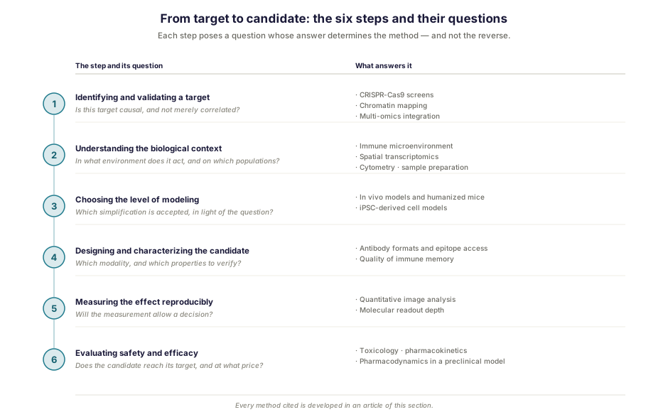
A research program running from a target to a candidate is not a chain of techniques. It is a sequence of questions, each conditioning the next, and the answer to each determines the method to be used — never the other way round. Choosing a tool because it is available, then looking for the question it might solve, is the surest way to produce data without producing a decision.
This article offers a map of these questions, step by step, referring for each to the articles in this section that treat it in detail. It closes on an observation that building the whole made visible, and that holds at every level.
Identifying and validating a target
The question at this stage is not whether the target is associated with the phenotype, but whether it is causal. A protein overexpressed in a disease may be a driver, a passenger or a consequence, and nothing in a measurement of abundance settles the matter.
What settles it is perturbation: remove, inhibit or restore, then observe. CRISPR-Cas9 screens industrialize this logic by interrogating thousands of genes in parallel, with two formats and two readout regimes according to whether one is looking for genes whose loss harms or genes whose loss confers an advantage.
Regulation counts as much as sequence. A target can be functional without being mutated, if its expression is locked by the state of the chromatin, and lifting that lock then constitutes a strategy in itself. Finally, crossing several layers of information sheds light on what a single one does not show, the gap between layers often being more instructive than their agreement.
The pitfall of this step: a co-occurrence is not a causation, and no volume of descriptive data makes up for it.
Understanding the biological context
A target does not act in a vacuum. The question becomes: in what environment, and on which cell populations?
In oncology, the composition of the immune infiltrate and above all its spatial organization carry prognostic information that abundance alone does not give — the same density of lymphocytes means something different according to whether they penetrate the tumor or remain at its periphery. When position counts, spatial transcriptomics measures expression without erasing the geography.
Phenotyping by cytometry identifies and quantifies populations, provided the panel was designed for the question posed. And upstream of any molecular measurement, sample preparation decides what one will be able to see: a fragile population lost at that step will reappear in no analysis.
The pitfall of this step: an average computed on a heterogeneous reality erases its relief.
Choosing the level of modeling
No model reproduces human disease. The question is therefore not to find the best one, but to know which simplification one accepts in light of the question posed.
In vivo models integrate the whole organism (circulation, immune system, interactions between organs) at the cost of differences between species, which genetic humanization sometimes allows to be circumvented by replacing a murine protein with its human homolog. Cell models derived from induced pluripotent stem cells offer the reverse: human material carrying the patient’s mutation, differentiable toward the affected cell type, but without systemic context. Their major methodological asset is the isogenic control, which allows a phenotype to be ascribed to a variant rather than to a genetic background. Where the object of study must be built before the question can be asked, another set of constraints applies.
The pitfall of this step: retaining the model available rather than the model relevant, then interpreting as though one had chosen the second.
Designing and characterizing the candidate
The question becomes one of modality and of the properties to be verified.
Format conditions access to the target. Single-domain antibodies, by their small size and the geometry of their binding site, reach buried epitopes that a flat binding site reaches with difficulty — a property that makes them useful as research tools and diagnostic reagents as much as therapeutic candidates.
For vaccine and immunotherapeutic approaches, the question of measurement shifts. An antibody titer describes a state at a given moment; the quality of the immune memory generated — diversity of clones, degree of maturation, breadth of variant recognition — tells more about durable protection, and is not obtained by serology.
The pitfall of this step: measuring what is easy rather than what predicts.
Measuring the effect reproducibly
That leaves the question that decides everything: will the measurement allow a conclusion?
Standardized image quantification turns an appraisal into a measurement, which matters particularly when a threshold separates two therapeutic courses. And the choice of molecular readout depth — an average profile across the whole sample, or a profile per cell — determines whether one will see a heterogeneous response or its average. The metabolome, for its part, offers the layer closest to the phenotype, provided what is expected of it is calibrated on the fraction that can actually be named.
The pitfall of this step: confusing accuracy with reproducibility. A threshold-based decision requires both, and it is the second that is neglected.
Evaluating safety and efficacy
Finally comes the step that decides passage into the clinic, and it poses three questions it would be imprudent to conflate: does the candidate reach its target at an administrable dose, does it produce the intended effect there, and at what price in terms of tolerability?
In vitro and in vivo studies of toxicology, pharmacokinetics and pharmacodynamics answer these questions in preclinical models, which refers directly back to the choice of modeling level made above, since the scope of an evaluation remains bounded by the relevance of the model on which it is conducted.
The pitfall of this step: concluding that something is ineffective without having measured exposure. Without pharmacokinetic data, a negative result does not allow a candidate that does not act to be distinguished from a candidate that never reached its target.
What recurs at every step
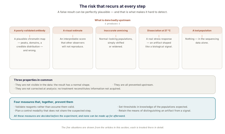
In building this section, one and the same risk appeared at every level, in different forms. It is the hardest to detect: a false result can be perfectly plausible.
An antibody that recognizes its target poorly produces a plausible chromatin map (peaks, domains, a credible distribution) that is nonetheless wrong; nothing in the data signals it. A semi-quantitative visual estimate of cell density produces an interpretable score that other observers will not reproduce. Spectral unmixing based on inaccurate reference signatures arrives at a result with no apparent error, simply shifting the populations. Enzymatic dissociation induces in the cells a transcriptional stress response — an artifact that has the shape of a biological signal, since it is one, merely triggered by the treatment. And a population entirely lost at preparation does not announce itself in the sequencing data: only an independent measurement can reveal its absence.
These five errors have three properties in common. They are not visible in the data, since they produce results of normal shape. They are not corrected at analysis, no statistical treatment reconstituting information that was not acquired. And they are all prevented upstream, by the same means: validating reagents rather than assuming them valid, planning a control modality that does not share the suspected step, setting thresholds in knowledge of the populations expected, and retaining the means of distinguishing an artifact from a signal.
It is there, more than in execution, that the value of methodological support is decided: knowing where the result may be wrong without looking wrong.
How Inovarion can support you
Inovarion works at each of these steps — target validation, characterization of the biological context, preclinical and cell models, candidate characterization, quantification and analysis. The value of a CRO lies less in executing an isolated technique than in the articulation of these steps: choosing the method that answers the question posed, and designing each step in knowledge of what the previous one made possible or definitively lost.
The articles in this section
Identifying and validating a target
- CRISPR-Cas9 screens: principles, strategies and applications
- Mapping chromatin: from ChIP-seq to CUT&Tag
- Multi-omics integration
Understanding the biological context
- Characterizing the tumor immune microenvironment
- Spatial transcriptomics: principle, limits and applications in neuroinflammation and myositis
- Designing a cytometry panel: spectral vs conventional
- Sample preparation for scRNA-seq
Choosing the level of modeling
- Humanized mouse models and in vivo preclinical models: an overview
- iPSC-derived cell models: principle, contributions and limits
- Synthetic biology
Designing and characterizing the candidate
- Single-domain antibodies (VHH) in hemostasis: research, diagnosis, therapy
- B cell memory and vaccine response: what single-cell adds
Measuring the effect reproducibly
- Digital pathology and quantitative image analysis
- scRNA-seq vs bulk RNA-seq: how to choose
- Metabolomics
Evaluating safety and efficacy
updated July 2026